
Structure Gallery
Biomolecular structures of BCIV staff
Below you will find all biomolecular structures determined by scientists of the Chair of Biochemistry IV by NMR, X-ray crystallography, cryo-EM or hybrid methods. For detailed information and to access the structure coordinates click the link to the protein data bank (PDB) or to the publication.
 |
|
|
|
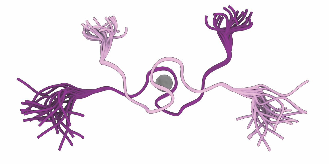
Solution structure of homodimeric TMEM106B PDB: 9GI8 Publication: Perez-Borrajero et al., EMBO Reports, 2026 |
||
 |
|
|
|
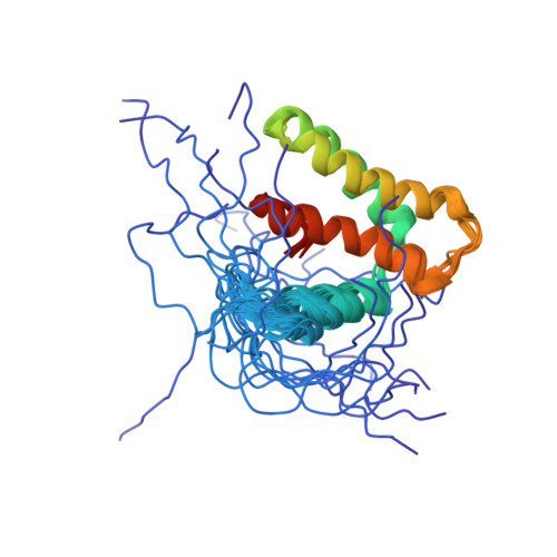
Solution structure of X55, a computationally designed protein PDB: 9HSA Publication: Jendrusch et al., Mol Syst Biol, 2025 |
||
 |
|
|
|
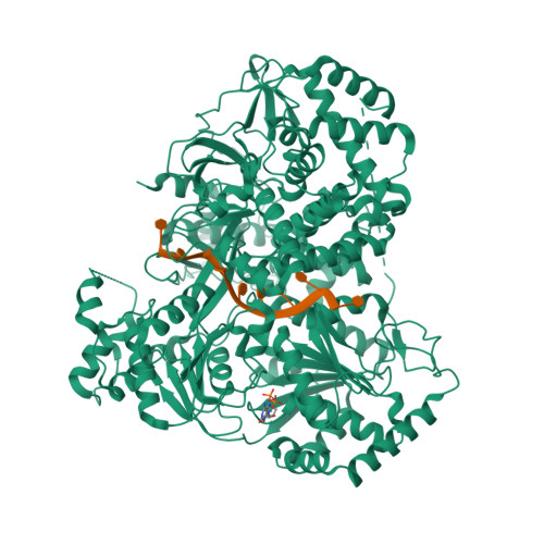
Cryo-EM structure of MLE in complex with UUC RNA and ADP PDB: 8PJB Publication: Jagtap et al., Mol Cell, 2023 |
||
 |
|
|
|
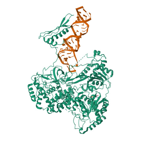
Cryo-EM structure of MLE in complex with SL7UUC RNA and ADP PDB: 8PJJ Publication: Jagtap et al., Mol Cell, 2023 |
||
 |
|
|
|
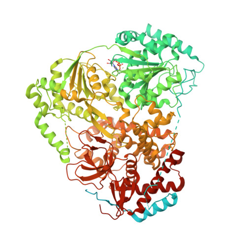
Cryo-EM structure of MLE in complex with ADP:AlF4 PDB: 8B9J Publication: Jagtap et al., Mol Cell, 2023 |
||
 |
|
|
|
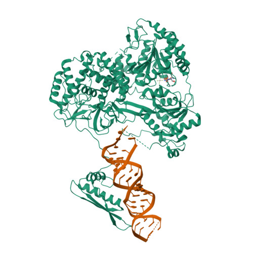
Cryo-EM structure of MLE in complex with ADP:AlF4 and SL7modUUC RNA PDB: 8B9K Publication: Jagtap et al., Mol Cell, 2023 |
||
 |
|
|
|
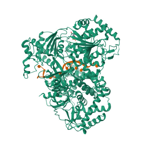
Cryo-EM structure of MLE in complex with ADP:AlF4 and UUC RNA PDB: 8B9I Publication: Jagtap et al., Mol Cell, 2023 |
||
 |
|
|
|
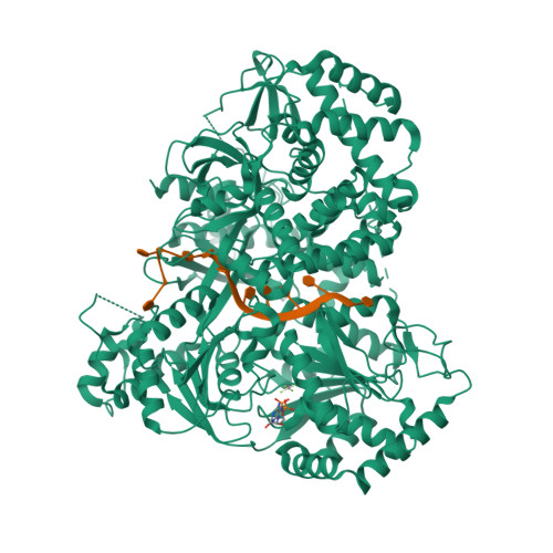
Cryo-EM structure of MLE in complex with ADP:AlF4 and U10 RNA PDB: 8B9G Publication: Jagtap et al., Mol Cell, 2023 |
||
 |
|
|
|
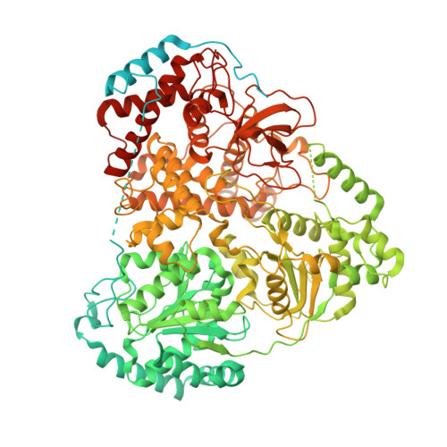
Cryo-EM structure of MLE PDB: 8B9L Publication: Jagtap et al., Mol Cell, 2023 |
||
 |
|
|
|
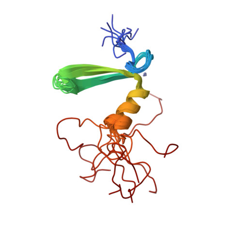
NMR structure of the N domain of AAA+ disaggregase ClpG PDB: 8P66 Publication: Katikaridis et al., J Biol Chen, 2023 |
||
 |
|
|
|
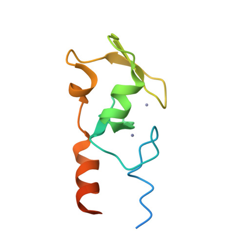
Crystal structure of the RING domain of human TRIM2 PDB: 8A38 Publication: Unpublished |
||
 |
|
|
|
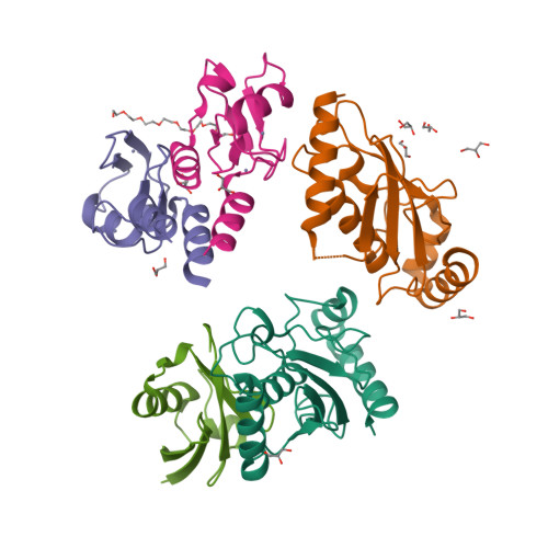
Crystal structure of the human TRIM2 RING, UBCH5C and ubiquitin complex PDB: 8AMS Publication: Unpublished |
||
 |
|
|
|
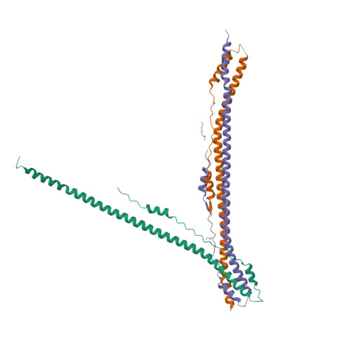
Crystal structure of the coiled-coil domain of human TRIM3 PDB: 8AMR Publication: Unpublished |
||
 |
|
|
|
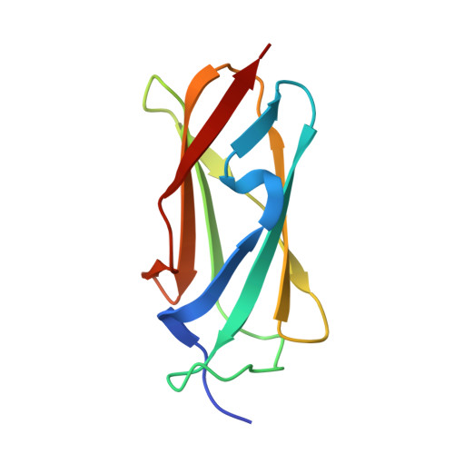
Crystal structure of the Filamin domain of human TRIM3 PDB: 7O0B Publication: Unpublished |
||
 |
|
|
|
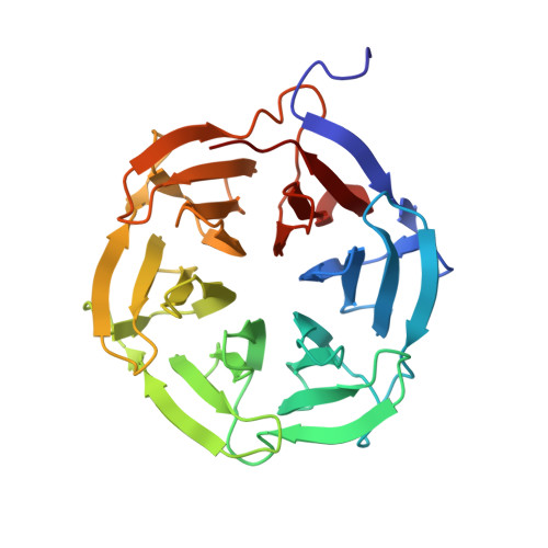
Crystal structure of the NHL domain of human TRIM2 PDB: 7B96 Publication: Unpublished |
||
 |
|
|
|
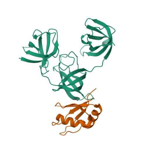
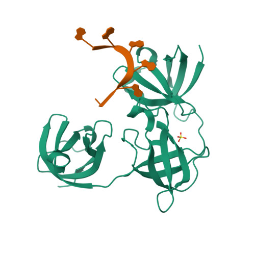
Crystal structure of the Drosophila Unr-CSD789:pAbp-RRM3 complex PDB: 7ZHR Publication: Hollmann et al., Nucleic Acids Res, 2023 |
||
 |
|
|
|
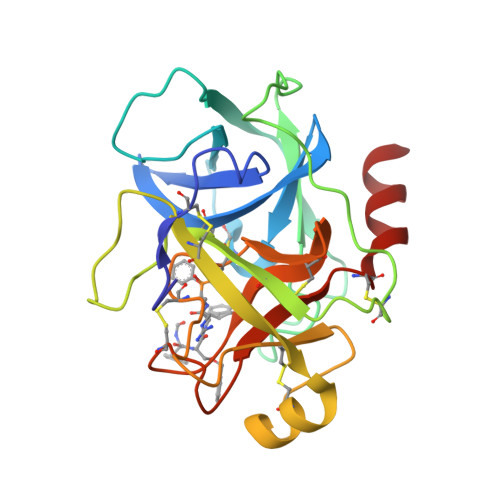
Crystal structure of the Drosophila Unr-CSD789:poly(A)-RNA complex PDB: 7ZHH Publication: Hollmann et al., Nucleic Acids Res, 2023 |
||
 |
|
|
|
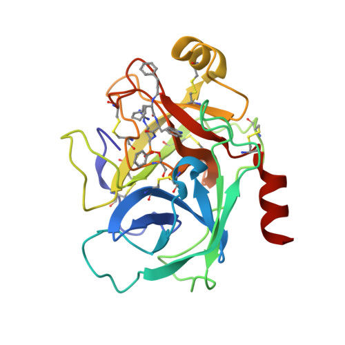
Crystal structure of KLK6 in complex with compound 17a PDB: 7QFV Publication: Zhang et al., JACS, 2022 |
||
 |
|
|
|
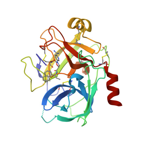
Crystal structure of KLK6 in complex with compound 16a PDB: 7QFT Publication: Zhang et al., JACS, 2022 |
||
 |
|
|
|
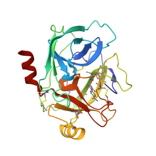
Crystal structure of KLK6 in complex with compound DKFZ918 PDB: 7QI0 Publication: Baumann et al., RSC Adv, 2022 |
||
 |
|
|
|
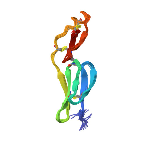
Crystal structure of KLK6 in complex with compound DKFZ917 PDB: 7QHZ Publication: Baumann et al., RSC Adv, 2022 |
||
 |
|
|
|
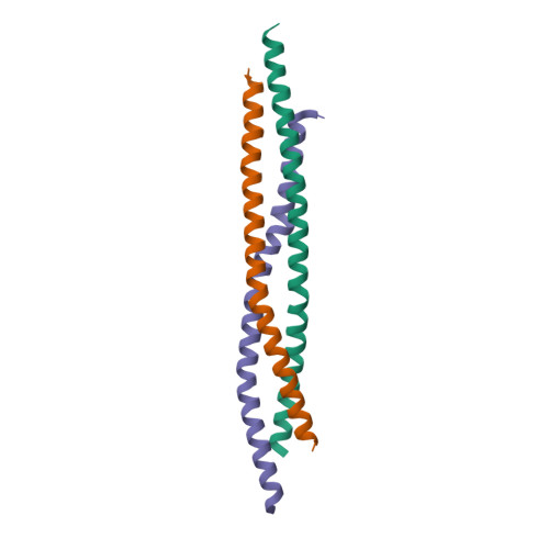
NMR structure of the C6 domain of von Willebrand Factor PDB: 7P4N Publication: Chen et al., J Struct Biol, 2022 |
||
 |
|
|
|
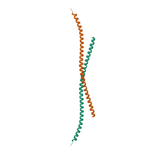
Crystal structure of the Khc/atypical Tropomyosin 1 complex PDB: 7BJS Publication: Dimitrova-Paternoga et al., Genes Dev, 2021 |
||
 |
|
|
|
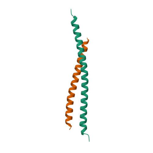
Crystal structure of atypical Tropomyosin 1, residues 262-363 PDB: 7BJG Publication: Dimitrova-Paternoga et al., Genes Dev, 2021 |
||
 |
|
|
|
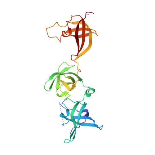
Crystal structure of atypical Tropomyosin 1, residues 270-334 PDB: 7BJN Publication: Dimitrova-Paternoga et al., Genes Dev, 2021 |
||
 |
|
|
|
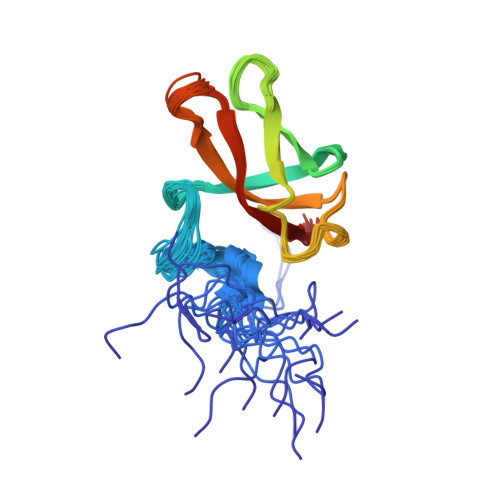
Crystal structure of Unr-CSD456 PDB: 6Y6E Publication: Hollmann et al., Cell Rep, 2020 |
||
 |
|
|
|
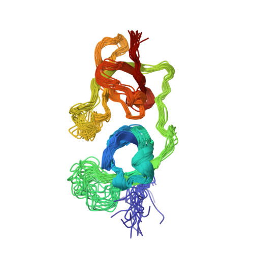
NMR structure of Unr-CSD9 PDB: 6Y96 Publication: Hollmann et al., Cell Rep, 2020 |
||
 |
|
|
|
NMR structure of Unr-CSD78 PDB: 6Y4H Publication: Hollmann et al., Cell Rep, 2020 |
||
 |
|
|
|
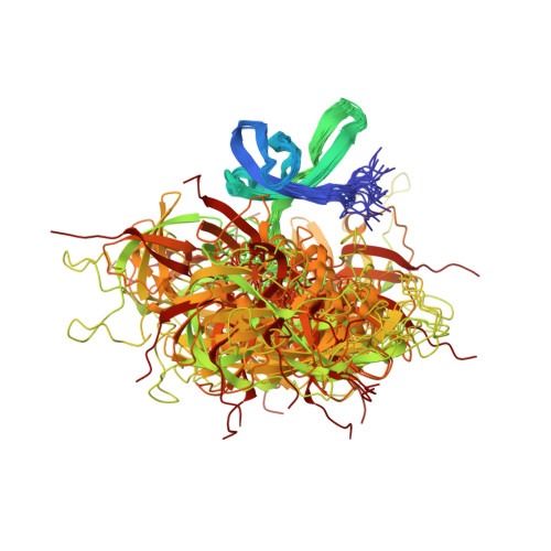
NMR structure of Unr-CSD12 PDB: 6Y6M Publication: Hollmann et al., Cell Rep, 2020 |
||
 |
|
|
|
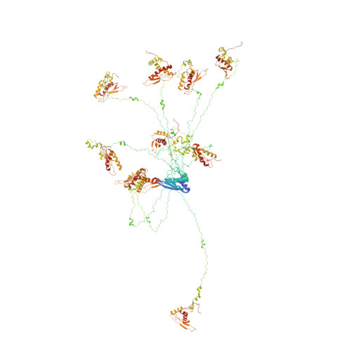
NMR structure of MLE-dsRBD12 PDB: 6I3R Publication: Jagtap et al., Nucleic Acids Res, 2019 |
||
 |
|
|
|
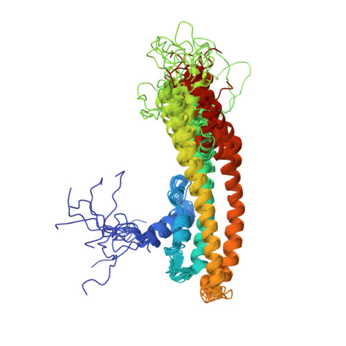
NMR structure of a part of Condensin in complex with Brn1 PDB: 6Q6E Publication: Hassler et al., Mol Cell, 2019 |
||
 |
|
|
|
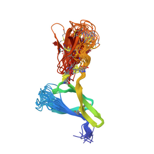
NMR structure of the C4 domain of von Willebrand Factor PDB: 6FWN Publication: Xu et al., Blood, 2019 |
||
 |
|
|
|
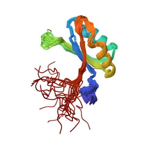
NMR structure of TIA-1-RRM1 PDB: 5O2V Publication: Sonntag et al., Angewandte Chemie Intl Ed, 2017 |
||
 |
|
|
|
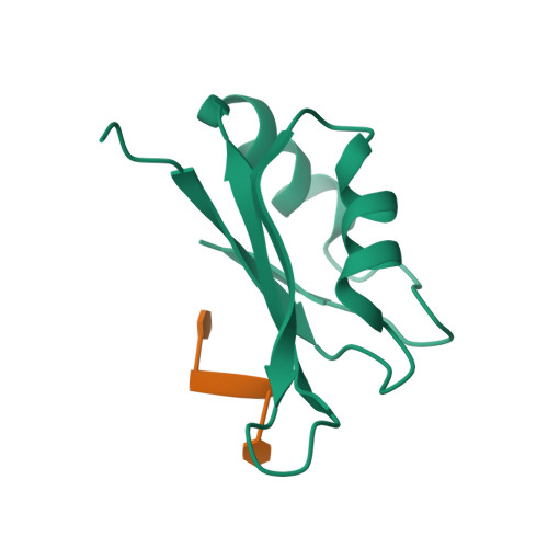
Crystal structure of TIA-1-RRM2 bound to RNA PDB: 5O3J Publication: Sonntag et al., Angewandte Chemie Intl Ed, 2017 |
||
 |
|
|
|
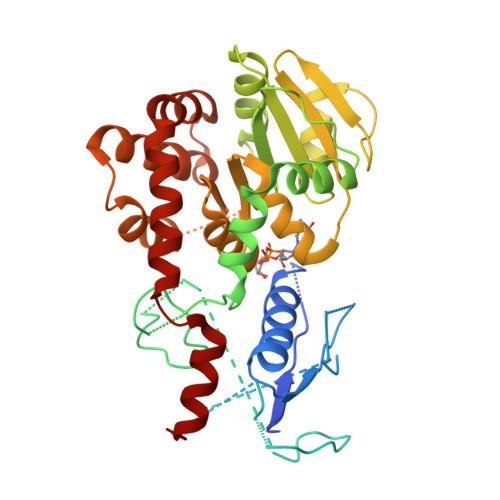
Crystal structure of the Rhamnosyl-transferase EarP PDB: 5NV8 Publication: Krafczyk et al., mBio, 2017 |
||
 |
|
|
|
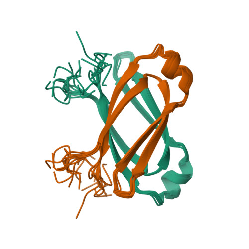
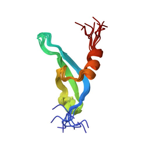
NMR structure of the EBNA-2 dimerization domain from the Epstein-Barr virus PDB: 2N2J Publication: Friberg et al., Plos Pathogens, 2015 |
||
 |
|
|
|
NMR structure of the chromodomain 3 of cpSRP43 PDB: 2N88 Publication: Horn et al., Nature Commun, 2015 |
||
 |
|
|
|
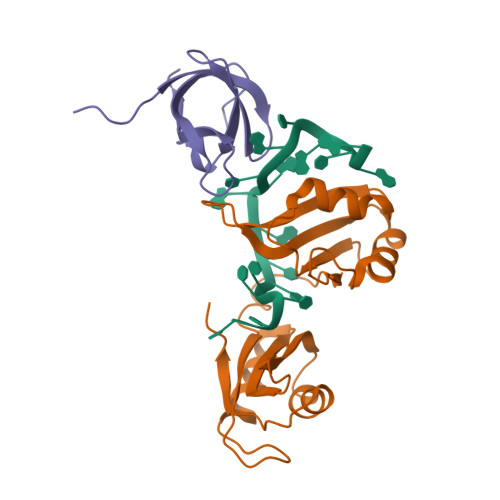
Crystal structure of the Sxl-Unr-msl2 mRNA complex PDB: 4QQB Publication: Hennig et al., Nature, 2014 |
||
 |
|
|
|
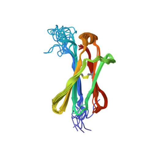
NMR structure of the fourth constant immunoglobulin domain of nurse shark igNAR PDB: 2MKL Publication: Feige et al., PNAS, 2014 |
||